The Totary-Jain Lab

Exploring the role of non-coding RNAs in immunity and the progression of atherosclerosis.


Atherosclerotic Cardiovascular Disease

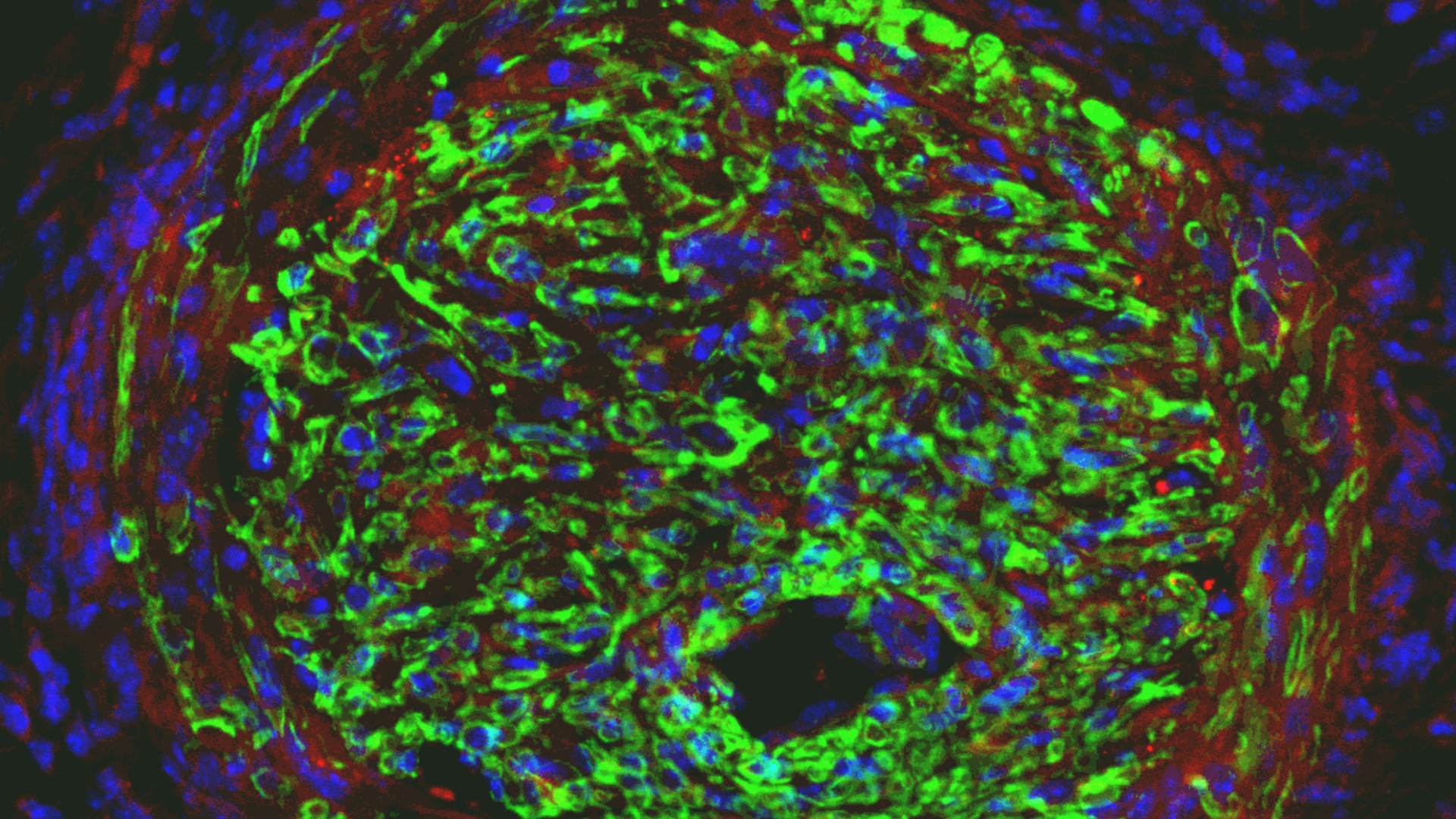
Endothelial cell coverage of balloon
injured arteries treated with cell selective
Ad-p27-126Ts miRNA switch
En-Face Endothelial cell staining of artery
exhibiting restenosis after balloon injury

Our lab aimed to develop cell-selective nanotherapy to combat atherosclerotic cardiovascular disease. This therapy specifically targets inflammatory and vascular smooth muscle cells, which are key players in plaque buildup, while carefully preserving healthy endothelial cells.

Initially, we employed an adenoviral vector, but our current focus is on a more advanced synthetic mRNA-based miRNA switch technology encapsulated in nanoparticles.

This innovative approach effectively delivers therapeutic payloads directly to damaged vessel areas, significantly reducing plaque formation and promoting vessel repair. We are actively enhancing this technology by combining it with siRNA to further suppress inflammation, in an effort to restore the endothelial barrier while inhibiting the progression of atherosclerotic plaque.

Transposable Elements and Antiviral Defense

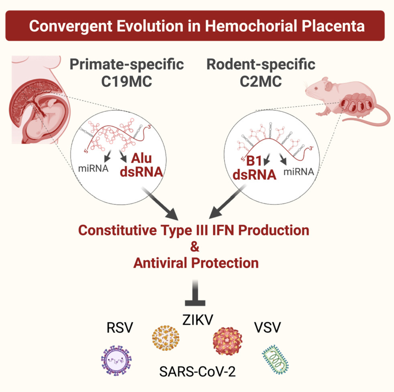
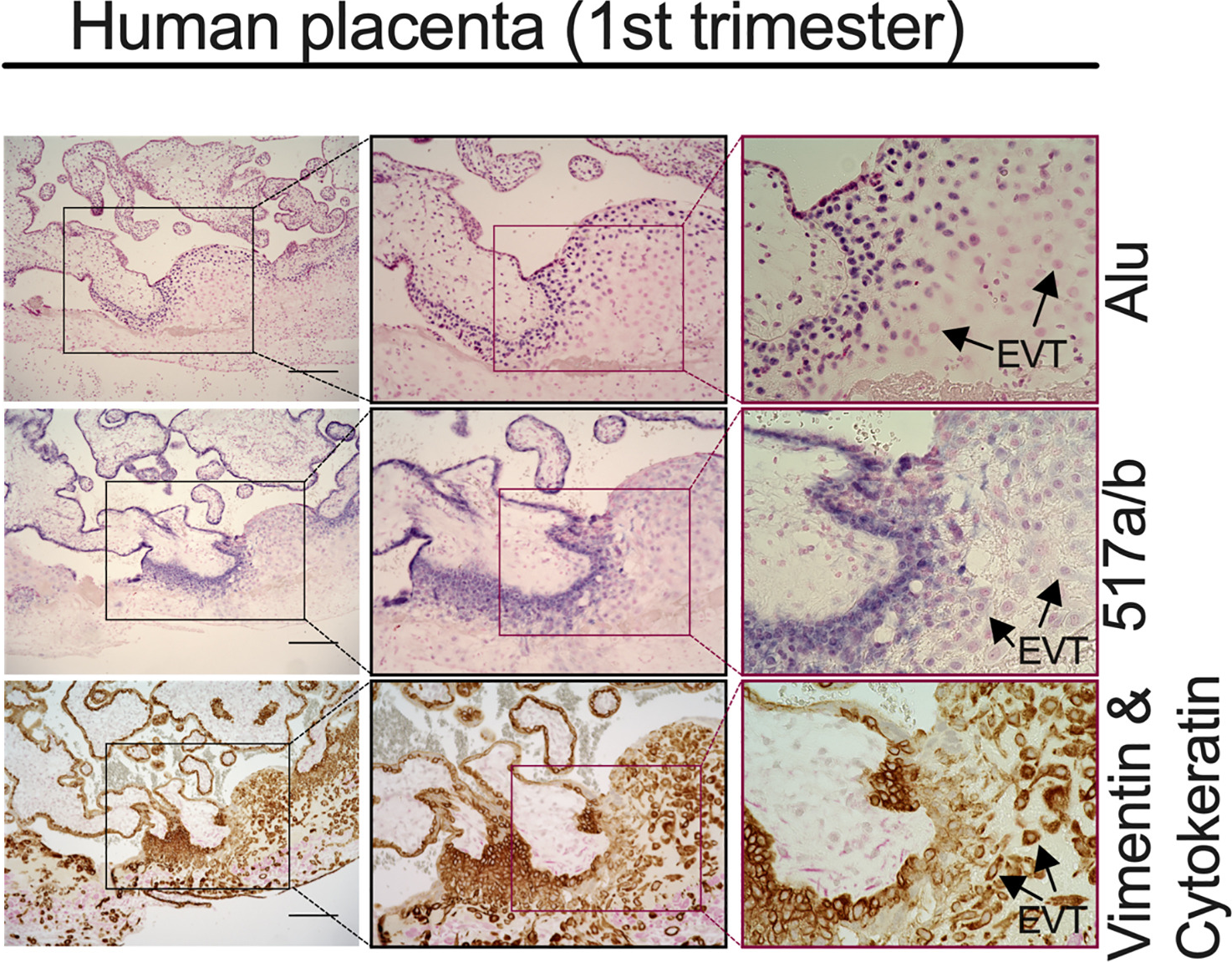
Our laboratory was the first to use CRISPR technology to transcriptionally activate the C19MC, a primate-specific microRNA cluster located on chromosome 19. This breakthrough enabled us to uncover a compelling example of convergent evolution involving the C19MC and a rodent-specific microRNA cluster on chromosome 2, C2MC. These clusters are primarily expressed in trophoblasts and are enriched with short interspersed nuclear elements (SINEs) known as Alu (primate) or B1 (rodent) repeats.

Our research has revealed that the SINEs of these clusters produce double-stranded RNA (dsRNA), which triggers a potent immune response akin to viral mimicry. This immune activation involves type III interferon signaling, providing crucial antiviral protection to the fetus during development. Furthermore, we also demonstrated that C19MC plays a pivotal role in regulating epithelial-to-mesenchymal transition and accelerating cell reprogramming processes.

Relevant Publications

  • de Assis, Viviana, Umit A Kayisli, Asli Ozmen, Nihan Semerci, Hana Totary-Jain, Nagehan Pakasticali, Ganesh Halade V, Charles J Lockwood, and Ozlem Guzeloglu-Kayisli. (2024) 2024. “Decidual Cells Block Inflammation-Mediated Inhibition of 15-Hydroxyprostaglandin Dehydrogenase in Trophoblasts.”. The American Journal of Pathology. https://doi.org/10.1016/j.ajpath.2024.05.005.

    Chorioamnionitis generates prostaglandin (PG) E2 and F2α, promoting fetal membrane rupture, cervical ripening, and uterine contractions. 15-Hydroxyprostaglandin dehydrogenase (HPGD) contributes to pregnancy maintenance by inactivating PGs. The role of decidual cells in regulating HPGD expression at the maternal-fetal interface was investigated. HPGD immunostaining was primarily detected in anchoring villi and choriodecidual extravillous trophoblasts (EVTs) in the first, second, and third trimesters. Chorionic EVTs adjacent to decidua parietalis exhibited significantly higher HPDG levels than those adjacent to amnion. HPGD histologic score levels were significantly lower in choriodecidua from chorioamnionitis versus gestational age-matched controls (means ± SEM, 132.6 ± 3.8 versus 31.2 ± 7.9; P < 0.05). Conditioned media supernatant (CMS) from in vitro decidualized term decidual cells (TDCs) up-regulated HPGD levels in EVTs differentiated from human trophoblastic stem cells, primary trophoblasts, and HTR8/SVneo cells. However, CMS from 5 μg/mL lipopolysaccharide or 10 ng/mL IL-1β pretreated TDC cultures down-regulated HPGD levels in HTR8/SVneo cultures. Similarly, direct treatment of HTR8/SVneo cultures with lipopolysaccharide or IL-1β significantly reduced HPGD levels versus control (0.57 ± 0.1 or 0.47 ± 0.1 versus 1.03 ± 0.03; P < 0.05) but not in TDC-CMS pretreated HTR8/SVneo cultures. Collectively, the results uncover a novel decidual cell-mediated paracrine mechanism that stimulates levels of trophoblastic HPGD, whose function is to inactivate labor-inducing PGs, thereby promoting uterine quiescence during pregnancy. However, infectious/inflammatory stimuli in decidual cells cause a paracrine inhibition of trophoblastic HPGD expression, increasing PGE2/PGF2α levels, thereby contributing to preterm birth.

  • Hetherington, Isabella, and Hana Totary-Jain. (2022) 2022. “Anti-Atherosclerotic Therapies: Milestones, Challenges, and Emerging Innovations.”. Molecular Therapy : The Journal of the American Society of Gene Therapy 30 (10): 3106-17. https://doi.org/10.1016/j.ymthe.2022.08.024.

    Atherosclerosis is the main underlying pathology for many cardiovascular diseases (CVDs), which are the leading cause of death globally and represent a serious health crisis. Atherosclerosis is a chronic condition that can lead to myocardial infarction, ischemic cardiomyopathy, stroke, and peripheral arterial disease. Elevated plasma lipids, hypertension, and high glucose are the major risk factors for developing atherosclerotic plaques. To date, most pharmacological therapies aim to control these risk factors, but they do not target the plaque-causing cells themselves. In patients with acute coronary syndromes, surgical revascularization with percutaneous coronary intervention has greatly reduced mortality rates. However, stent thrombosis and neo-atherosclerosis have emerged as major safety concerns of drug eluting stents due to delayed re-endothelialization. This review summarizes the major milestones, strengths, and limitations of current anti-atherosclerotic therapies. It provides an overview of the recent discoveries and emerging game-changing technologies in the fields of nanomedicine, mRNA therapeutics, and gene editing that have the potential to revolutionize CVD clinical practice by steering it toward precision medicine.

  • Ozmen, Asli, Ozlem Guzeloglu-Kayisli, Selcuk Tabak, Xiaofang Guo, Nihan Semerci, Chinedu Nwabuobi, Kellie Larsen, et al. (2022) 2022. “Preeclampsia Is Associated With Reduced ISG15 Levels Impairing Extravillous Trophoblast Invasion.”. Frontiers in Cell and Developmental Biology 10: 898088. https://doi.org/10.3389/fcell.2022.898088.

    Among several interleukin (IL)-6 family members, only IL-6 and IL-11 require a gp130 protein homodimer for intracellular signaling due to lack of intracellular signaling domain in the IL-6 receptor (IL-6R) and IL-11R. We previously reported enhanced decidual IL-6 and IL-11 levels at the maternal-fetal interface with significantly higher peri-membranous IL-6 immunostaining in adjacent interstitial trophoblasts in preeclampsia (PE) vs. gestational age (GA)-matched controls. This led us to hypothesize that competitive binding of these cytokines to the gp130 impairs extravillous trophoblast (EVT) differentiation, proliferation and/or invasion. Using global microarray analysis, the current study identified inhibition of interferon-stimulated gene 15 (ISG15) as the only gene affected by both IL-6 plus IL-11 vs. control or IL-6 or IL-11 treatment of primary human cytotrophoblast cultures. ISG15 immunostaining was specific to EVTs among other trophoblast types in the first and third trimester placental specimens, and significantly lower ISG15 levels were observed in EVT from PE vs. GA-matched control placentae (p = 0.006). Induction of primary trophoblastic stem cell cultures toward EVT linage increased ISG15 mRNA levels by 7.8-fold (p = 0.004). ISG15 silencing in HTR8/SVneo cultures, a first trimester EVT cell line, inhibited invasion, proliferation, expression of ITGB1 (a cell migration receptor) and filamentous actin while increasing expression of ITGB4 (a receptor for hemi-desmosomal adhesion). Moreover, ISG15 silencing further enhanced levels of IL-1β-induced pro-inflammatory cytokines (CXCL8, IL-6 and CCL2) in HTR8/SVneo cells. Collectively, these results indicate that ISG15 acts as a critical regulator of EVT morphology and function and that diminished ISG15 expression is associated with PE, potentially mediating reduced interstitial trophoblast invasion and enhancing local inflammation at the maternal-fetal interface. Thus, agents inducing ISG15 expression may provide a novel therapeutic approach in PE.