Publications

Human minibrains reveal effects of psychedelic substance

Publications

“Restoring sight through regenerative innovation.”


  • Du, Rong, Ajay Kumar, Enzhi Yang, Jingxue Zhang, Ningli Wang, and Yiqin Du. 2025. “Transcriptomic Differences Between Human Trabecular Meshwork Stem Cells and Trabecular Meshwork Cells Reveal Specific Biomarker Profiles”. Current Issues in Molecular Biology 47 (7). https://doi.org/10.3390/cimb47070514.
    Glaucoma is a leading cause of irreversible blindness, normally associated with dysfunction and degeneration of the trabecular meshwork (TM) as the primary cause. Trabecular meshwork stem cells (TMSCs) have emerged as promising candidates for TM regeneration toward glaucoma therapies, yet their molecular characteristics remain poorly defined. In this study, we performed a comprehensive transcriptomic comparison of human TMSCs and human TM cells (TMCs) using RNA sequencing and microarray analyses, followed by qPCR validation. A total of 465 differentially expressed genes were identified, with 254 upregulated in TMSCs and 211 in TMCs. A functional enrichment analysis revealed that TMSCs are associated with development, immune signaling, and extracellular matrix remodeling pathways, while TMCs are enriched in structural, contractile, and adhesion-related functions. A network topology analysis identified CXCL3, CXCL6, and BMP2 as robust TMSC-specific hub genes, and LMOD1 and BGN as TMC-specific markers, with expression patterns confirmed by qPCR. These findings define distinct molecular signatures of TMSCs and TMCs, providing reliable biomarkers for cell identity and a foundation for future stem cell-based therapies targeting TM dysfunction in glaucoma.
  • Du, Rong, Enzhi Yang, Madison Clark, Ningli Wang, and Yiqin Du. (2025) 2025. “Identification and Validation of Key Biomarkers in the Proximal Aqueous Humor Outflow Pathway.”. Current Issues in Molecular Biology 47 (3). https://doi.org/10.3390/cimb47030147.

    Glaucoma is a leading cause of irreversible blindness, with elevated intraocular pressure (IOP) as the most important risk factor. The trabecular meshwork (TM) and Schlemm's canal are the main components of the proximal aqueous humor outflow pathway. Their dysfunction is a major contributor to IOP elevation. This study aims to identify and validate key biomarkers for TM and Schlemm's canal endothelial (SCE) cells. A Microarray was performed on characterized human TM and SCE cells to analyze their transcriptome profiling. Differentially expressed genes (DEGs) were identified and cross-referenced with published single-cell RNA sequencing (scRNA-Seq) datasets to ensure cell-specific relevance. Further validation was performed using qPCR and re-confirmed on the scRNA-seq datasets. One-way ANOVA was used for statistical analysis, and p < 0.05 was considered significant. The Microarray revealed 341 DEGs, with TM cells enriched in metabolic and signaling pathways and SCE cells enriched in adhesion, immune, and morphogenesis-related processes. Cross-referencing with scRNA-Seq data refined the list of candidate biomarkers, and qPCR confirmed the significant gene expression differences between TM and SCE cells. CTTNBP2 and MGARP were identified as TM cell markers. JAM2, PODXL, and IFI27 are new SCE cell biomarkers. The validated biomarkers offer insights into glaucoma pathophysiology and lay the groundwork for targeted therapies.

  • Kumar, Ajay, Enzhi Yang, and Yiqin Du. (2025) 2025. “Trabecular Meshwork Regeneration for Glaucoma Treatment Using Stem Cell-Derived Trophic Factors.”. Methods in Molecular Biology (Clifton, N.J.) 2848: 59-71. https://doi.org/10.1007/978-1-0716-4087-6_4.

    Glaucoma is one of the leading causes of irreversible blindness. Stem cell therapy has shown promise in the treatment of primary open-angle glaucoma in animal models. Stem cell-free therapy using stem cell-derived trophic factors might be in demand in patients with high-risk conditions or religious restrictions. In this chapter, we describe methods for trabecular meshwork stem cell (TMSC) cultivation, secretome harvesting, and protein isolation, as well as assays to ensure the health of TMSC post-secretome harvesting and for secretome periocular injection into mice for therapeutic purposes.

  • Du, Yiqin, Sridhar Bammidi, and Enzhi Yang. (2025) 2025. “Trabecular Meshwork Stem Cells for Glaucoma Treatment.”. Methods in Molecular Biology (Clifton, N.J.) 2858: 143-58. https://doi.org/10.1007/978-1-0716-4140-8_13.

    Elevated intraocular pressure (IOP) is the most important risk factor for primary open-angle glaucoma (POAG) and currently is the only effective treatment target for glaucoma to prevent vision loss. In POAG patients, the trabecular meshwork (TM) cellularity is reduced which might be the main pathologic reason for the conventional outflow pathway dysfunction leading to elevated IOP. Stem cell-based therapy has been shown promising to reduce IOP and preserve retinal ganglion cells and their function in animal models. In this chapter, we describe the method details on TM stem cell cultivation and identification; induction for differentiation into different cell types, including differentiation to TM cell responsiveness to dexamethasone treatment with phagocytic function; and transplantation into mouse anterior chamber for therapeutic purposes.

  • Santra, Mithun, Moira L Geary, Elizabeth Rubin, Michael Y S Hsu, Martha L Funderburgh, Christine Chandran, Yiqin Du, Deepinder K Dhaliwal, Vishal Jhanji, and Gary Hin-Fai Yam. (2024) 2024. “Good Manufacturing Practice Production of Human Corneal Limbus-Derived Stromal Stem Cells and in Vitro Quality Screening for Therapeutic Inhibition of Corneal Scarring.”. Stem Cell Research & Therapy 15 (1): 11. https://doi.org/10.1186/s13287-023-03626-8.

    BACKGROUND: Mesenchymal stem cells in the adult corneal stroma (named corneal stromal stem cells, CSSCs) inhibit corneal inflammation and scarring and restore corneal clarity in pre-clinical corneal injury models. This cell therapy could alleviate the heavy reliance on donor materials for corneal transplantation to treat corneal opacities. Herein, we established Good Manufacturing Practice (GMP) protocols for CSSC isolation, propagation, and cryostorage, and developed in vitro quality control (QC) metric for in vivo anti-scarring potency of CSSCs in treating corneal opacities.

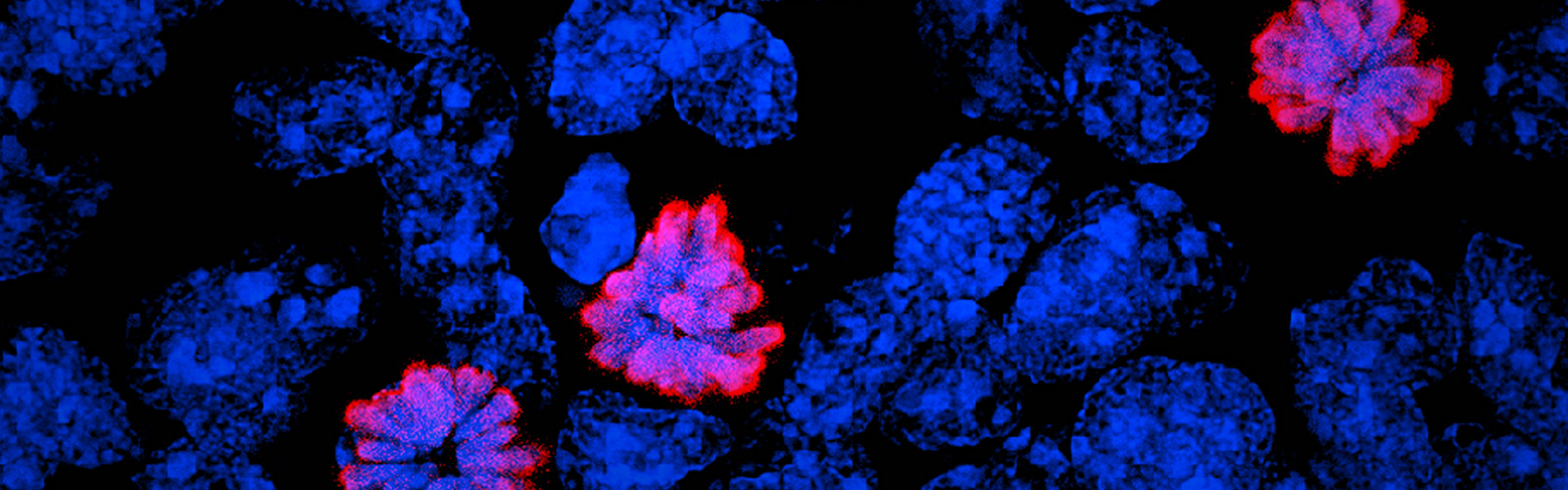
    METHODS: A total of 24 donor corneal rims with informed consent were used-18 were processed for the GMP optimization of CSSC culture and QC assay development, while CSSCs from the remaining 6 were raised under GMP-optimized conditions and used for QC validation. The cell viability, growth, substrate adhesion, stem cell phenotypes, and differentiation into stromal keratocytes were assayed by monitoring the electric impedance changes using xCELLigence real-time cell analyzer, quantitative PCR, and immunofluorescence. CSSC's conditioned media were tested for the anti-inflammatory activity using an osteoclastogenesis assay with mouse macrophage RAW264.7 cells. In vivo scar inhibitory outcomes were verified using a mouse model of anterior stromal injury caused by mechanical ablation using an Algerbrush burring.

    RESULTS: By comparatively assessing various GMP-compliant reagents with the corresponding non-GMP research-grade chemicals used in the laboratory-based protocols, we finalized GMP protocols covering donor limbal stromal tissue processing, enzymatic digestion, primary CSSC culture, and cryopreservation. In establishing the in vitro QC metric, two parameters-stemness stability of ABCG2 and nestin and anti-inflammatory ability (rate of inflammation)-were factored into a novel formula to calculate a Scarring Index (SI) for each CSSC batch. Correlating with the in vivo scar inhibitory outcomes, the CSSC batches with SI < 10 had a predicted 50% scar reduction potency, whereas cells with SI > 10 were ineffective to inhibit scarring.

    CONCLUSIONS: We established a full GMP-compliant protocol for donor CSSC cultivation, which is essential toward clinical-grade cell manufacturing. A novel in vitro QC-in vivo potency correlation was developed to predict the anti-scarring efficacy of donor CSSCs in treating corneal opacities. This method is applicable to other cell-based therapies and pharmacological treatments.

  • Acott, Ted S, Michael P Fautsch, Weiming Mao, Ross Ethier, Alex S Huang, Mary J Kelley, Mini Aga, et al. (2024) 2024. “Consensus Recommendations for Studies of Outflow Facility and Intraocular Pressure Regulation Using Ex Vivo Perfusion Approaches.”. Investigative Ophthalmology & Visual Science 65 (14): 32. https://doi.org/10.1167/iovs.65.14.32.

    Intraocular pressure (IOP) elevation is the primary risk factor and currently the main treatable factor for progression of glaucomatous optic neuropathy. In addition to direct clinical and living animal in vivo studies, ex vivo perfusion of anterior segments and whole eyes is a key technique for studying conventional outflow function as it is responsible for IOP regulation. We present well-tested experimental details, protocols, considerations, advantages, and limitations of several ex vivo model systems for studying IOP regulation. These include: (1) perfused whole globes, (2) stationary anterior segment organ culture, (3) perfused human anterior segment organ culture, (4) perfused animal anterior segment organ culture, (5) perfused human corneal rims, and (6) perfused human anterior segment wedges. These methods, with due consideration paid to their strengths and limitations, comprise a set of very strong tools for extending our understanding of IOP regulation.