Research

Slow-Release Drug Delivery

With our novel preparations of essential drugs in ophthalmology, we utilize unique polymer formulations and conjugation/encapsulation to develop slow-release drug release technologies.
Drug Discovery

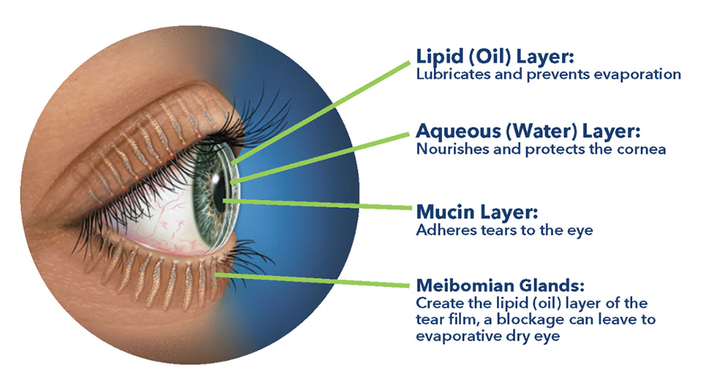
Our team is developing unique formulations for addressing dry eye disease and pan-antimicrobial solutions for all forms of keratitis (bacterial, fungal, and viral).


Technology and Engineering

We aim to develop innovating applications for technology and engineering, bridging it with medicine to improve patient outcomes.

Recent Publications

  • Sahiner, Nurettin, Daniel J Kravitz, Rabah and Qadir, Diane A Blake, Salima Haque, Vijay T John, Curtis E Margo, and Ramesh S Ayyala. 2009. “Creation of a Drug-Coated Glaucoma Drainage Device Using Polymer Technology: In Vitro and in Vivo Studies”. Arch. Ophthalmol. 127 (4): 448-53.

    "OBJECTIVE: To create and test a slow-release antifibrotic drug-coated glaucoma drainage device using in vitro and in vivo experiments. METHODS: A slow-release device incorporating mitomycin C in poly(2-hydroxyethyl methacrylate) disks was developed using redox-polymerization techniques. A standardized preparation of this drug delivery device was attached to the Ahmed glaucoma valve (model FP7; New World Medical, Inc, Rancho Cucamonga, California). Semicircular disks (5 x 6 mm) of P(HEMA)-mitomycin C containing varying concentrations of mitomycin C per gram dry weight of the gel were attached to the lower half of an Ahmed glaucoma valve plate. Water was pumped through the modified Ahmed glaucoma valve at a rate comparable to that of aqueous humor outflow, and mitomycin C release was measured. Modified and unmodified Ahmed glaucoma valves were implanted in a rabbit model, and drug release and fibrosis were assessed after 3 months. RESULTS: The P(HEMA)-mitomycin C device released mitomycin C in vitro over 1 to 2 weeks. Studies in rabbits revealed that mitomycin C was released from the disks during the 3-month implantation. Histologic analysis demonstrated a significant reduction in inflammatory reaction and fibrosis in the resulting blebs. CONCLUSION: Our slow-release drug-coated glaucoma drainage device decreased fibrosis and inflammation in the resulting bleb in a rabbit model. CLINICAL RELEVANCE: This device could reduce the failure rate of glaucoma drainage devices."

  • Blake, Diane A ", Nurettin Sahiner, Vijay T and John, Antoine D Clinton, Korianne E Galler, Molly Walsh, Analisa Arosemena, Pamela Y Johnson, and Ramesh S Ayyala. 2006. “Inhibition of Cell Proliferation by Mitomycin C Incorporated into P(HEMA) Hydrogels”. J. Glaucoma 15 (4): 291-98.

    "OBJECTIVES: The technique of mitomycin C (MMC) drug delivery and its application in glaucoma surgery are not standardized with resultant inconsistencies in the results. Also, one time application of MMC does not seem to have the same efficacy after glaucoma drainage device surgeries compared with trabeculectomies. This preliminary study examined the efficacy of a slow release form of MMC for its ability to inhibit cell proliferation in vitro. METHODS: MMC was incorporated into 1% P(HEMA) hydrogels using a redox polymerization method. For some experiments, unreacted low molecular weight components were removed from the hydrogels before the MMC was incorporated. Sterile disks (8 mm) of each polymer sample were affixed to 60 mm tissue culture dishes, and the dishes were inoculated with COS-1 cells or early passage human conjunctival fibroblasts. After 7 days in culture, the number of cells in each dish was determined. Cell morphology was assessed in replicate cultures after fixation and staining. RESULTS: Hydrogels with unreacted low molecular weight components slowed cell proliferation and induced morphologic changes. Early passage human conjunctival fibroblasts were more sensitive than COS-1 cells both to intrinsic contaminants in the hydrogels and to incorporated MMC. Once contaminants had been removed, MMC-loaded hydrogels inhibited conjunctival fibroblast proliferation in a dose-dependent fashion, with an IC50 of approximately 0.15 mg/g polymer. CONCLUSIONS: This study demonstrates that a slow release form of MMC can inhibit cell proliferation in vitro. Future experiments will focus upon the efficacy of this polymer-bound form during in vivo wound healing."

  • Ayyala, Ramesh S, Jessica Laursen Duarte, and . 2006. “Glaucoma Drainage Devices: State of the Art”. Expert Rev. Med. Devices 3 (4): 509-21.

    "Glaucoma drainage devices (GDDs) create an alternate aqueous pathway by channeling aqueous from the anterior chamber through a long tube to an equatorial plate, inserted under the conjunctiva, which promotes bleb formation. GDDs are being used more frequently in the treatment of glaucoma, both as the primary procedure of choice and following failure of trabeculectomy operations. This article outlines the current concepts involving different GDDs, surgical techniques and a review of the current literature. In addition, the importance of the biomaterial and its implications for the success of the operation are discussed."

  • Suner, Selin S ", Sahin Demirci, Berkant and Yetiskin, Rawil Fakhrullin, Ekaterina Naumenko, Oguz Okay, Ramesh S Ayyala, and Nurettin " Sahiner. 2019. “Cryogel Composites Based on Hyaluronic Acid and Halloysite Nanotubes As Scaffold for Tissue Engineering”. Int. J. Biol. Macromol. 130: 627-35.

    "We present here preparation of mechanically strong and biocompatible cryogel composites based on hyaluronic acid (HA) and halloysite nanotubes (HNTs) of various compositions, and their applications as scaffold for different cell growing media. Uniaxial compression tests reveal that the incorporation of HNTs into HA cryogels leads to a  2.5-fold increase in their Young moduli, e.g., from 38 $\pm$ 1 to 99 $\pm$ 4 kPa at a HA:HNTs weight ratio of 1:2. Although HA:HNTs based cryogels were found to be blood compatible with 1.37 $\pm$ 0.11% hemolysis ratio at a HA:HNTs weight ratio of 1:2, they trigger thrombogenic activity with a blood clotting index of 17.3 $\pm$ 4.8. Remarkably, HA:HNTs cryogel composites were found to be excellent scaffold materials in the proliferation of rat mesenchymal stem cells (MSC), human cervical carcinoma cells (HeLa), and human colon cancer cells (HCT116). The cell studies revealed that an increased amount of HNT embedding into HA cryogels leads to an increase of MSC proliferation."

  • Sahiner, Nurettin ", Selin S Suner, and Ramesh S" Ayyala. 2019. “Mesoporous, Degradable Hyaluronic Acid Microparticles for Sustainable Drug Delivery Application”. Colloids Surf. B Biointerfaces 177: 284-93.

    "Porous and degradable hyaluronic acid (HA) microparticles was synthesized in a single step using different ratio of crosslinker, divinylsulfone (DVS) ranging between 2.5 and 100% mole ratio of HA repeating unit. HA particles less than 25% ($łeq$10%) crosslinker ratio were found to be mesoporous and provided the highest surface area, calculated as 21.54$\pm$10.31 m2/g for 2.5% crosslinked HA particles via BET analysis. Hydrolytic degradation of 2.5% crosslinked HA microparticles in PBS (pH 7.4) and at 37.5 ºC revealed a linear weight loss up to 20 days and 94.5$\pm$4.5% weight loss for 30 days was attained. A wide spectrum antibiotic, Vancomycin as a model drug was loaded to mesoporous HA particles via directly loading from aqueous corresponding solution and by chemical conjugation method to obtain controllable and sustained release profiles from HA particles. Up to 168 h linear vancomycin release (50.5$\pm$4.2 mg/g) was accomplished from 2.5% DVS crosslinked HA particles."

  • Sahiner, Nurettin ", Selin Sagbas, Mehtap and Sahiner, and Ramesh S Ayyala. 2017. “Polyethyleneimine Modified Poly(Hyaluronic Acid) Particles With Controllable Antimicrobial and Anticancer Effects”. Carbohydr. Polym. 159: 29-38.

    "Poly(hyaluronic acid) (p(HA)) particles with sizes from few hundred nm to few tens of micrometer were synthesized by using epoxy groups containing crosslinker glycerol diglycidyl ether (GDE) with high yield, 94$\pm$5%. P(HA) particles were oxidized by treatment with sodium periodate and then reacted with cationic polyethyleneimine (PEI) at 1:0.5, 1:1, and 1:2 wt ratio of p(HA):PEI to obtain p(HA)-PEI particles. From zeta potential measurements, isoelectronic points of bare p(HA) particles increased to pH 8.7 from 2.7 after modification with cationic PEI. New properties, such as antibacterial property, were attained for p(HA)-PEI after modification. The highest minimum bactericidal concentration (MBC) values were 0.5, 1, and 0.5mg/mL against Escherichia coli, Staphylococcus aureus, and Bacillus subtilis species for 1:0.5 ratio of p(HA)-PEI at 72h incubation time. Moreover, the p(HA)-PEI particles were found to be biocompatible with L929 fibroblast cells, and interestingly, p(HA)-PEI particles were found to inhibit MDA-MB-231 breast and H1299 cancer cell growth depending on amount of PEI in p(HA)-PEI particles."